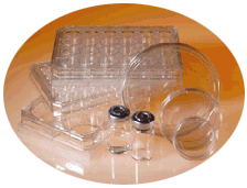
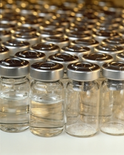
SmartPlastic: The Intelligent Choice for Enhanced Cell Performance SmartPlastic: Smart Products for Cell Culture


EMPOWER: Unleash the Power of the Extracellular Matrix

Extracellular matrix (ECM) proteins are an essential requirement for your cells to be productive. The matrix provides cells with physical support and dramatically influences their behavior. Basic cell functions are all related to -- and dependent on -- the matrix1.

By immediately binding to receptors on the cell surface, SmartPlastic® signals your cells that adhesion to a critical ECM protein sequence has occurred, influencing the survival, metabolism and fate of the cell, its shape, and many of its other properties. The sequence is from human fibronectin and contains the tripeptide RGD (Arg-Gly-Asp), also found within most major types of matrix proteins2.

SmartPlastic®presents the RGD cell attachment sequence directly on the culture surface in the form of ProNectin® F, a genetically engineered cell attachment factor. SmartPlastic®supports receptor-mediated cell adhesion and spreading for most anchorage-dependent cells, initiating cell growth.

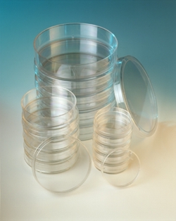
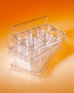
SmartPlastic: Tissue Culture Dishes SmartPlastic: Multi-Well Plates ProNectin Reagents for Cell Culture


ENHANCE: Improve Cell Productivity & Performance

"What can ProNectin F do for me?"

ProNectin® F customers report the following benefits in a variety of applications:
- more in vivo-like morphology 12,13,14,15
- faster and stronger cell adherence 3,4,5,7,8
- increased expression efficiency 9,10,11
Customers also report:
- improved plating efficiencies
- better growth

"What can ProNectin F do for me?"

ProNectin® F performance has been demonstrated with a variety of mammalian cell types including:

  • Bone cells
  • Embryonic cells
  • Endothelial cells
  • Epithelial cells
  • Eye-derived cells
  • Fibroblast cells
  • Glial cells
  • Hematopoietic cells
  • Muscle cells
  • Neuronal cells
  • Parenchymal cells
  • Tumor cells
  • A list of specific cell types is also available.


    "What can ProNectin F do for me?"

    ProNectin® F plays a vital role in a broad range of scientific applications -- in some cases enabling researchers to accomplish what might otherwise not have been possible. ProNectin® Fprovides a uniquely defined platform for applications in the fields of cell biology, cellular biochemistry, immunology, and developmental biology.

    "What can ProNectin F do for me?"

    ProNectin® F facilitates in vivo-like conditions in in vitro models:
    - Endothelial cells cultured under continuous flow12
    - Perfused transcapillary smooth muscle/endothelial co-culture13
    - Mammalian blood-brain barrier model14
    - CD34+ hematopoietic stem cell expansion in co-culture15

    Efficient Primary Cell Isolation
    Excellent viability achieved with primary cells on SmartPlastic® at a fraction of the time otherwise required7.

    Enhanced Production of Virus and Recombinant Proteins

    Increased DNA Transfection Efficiency
    ProNectin® F yielded DNA transfection efficiencies of adherent cell lines up to 20 times higher than standard tissue culture labware9.
    Improved Viral Infection Efficiency
    SmartPlastic® enabled fibroblast cultures to be infected with virus at 30% versus 50% of confluency10.
    Efficient Production of Recombinant Protein
    SmartPlastic® enabled serum-free production of gamma-interferon in recombinant CHO cells to reach level equivalent to 10% serum-containing media for up to 25 days11.

    Rapid Adhesion and Spreading of Aortic Endothelial Cells
    SmartPlastic® improved the adhesion and spreading of bovine aortic endothelial cells (BAEC) in serum-free media, compared to standard tissue culture labware. After 45 minutes, normal cobblestone morphology was apparent using SmartPlastic®3.

    Serum-Free BAEC: 45 Minutes Post-Seeding

    Serum-Free BAEC on standard tissue culture labware

    Serum-free BAEC on SmartPlastic with ProNectin F
    Standard Tissue Culture Labware SmartPlastic® with ProNectin® F


    ProNectin F at Work in Cellular Biochemistry and Developmental Biology

    • Investigation of in vitrocell-matrix adhesion in intact and non-lethally damaged renal tubular epithelial cells6
    • Demonstration of the role of the RGD cell-binding domain in trophoblast adhesion and possible impacts on in utero implantation16
    • Exploration of signal-dependent protein import in a permeabilized cell system8
    • Identification of mAb reactivity with specific integrins and receptor- related functionality17
    • Molecular immunology applications19,20
    • Demonstration of the beneficial effects of co-culture with cumulus cells in human in vitro fertilization24

    Rapid Adaptation of Serum Grown Cell Lines to Serum-Free Media
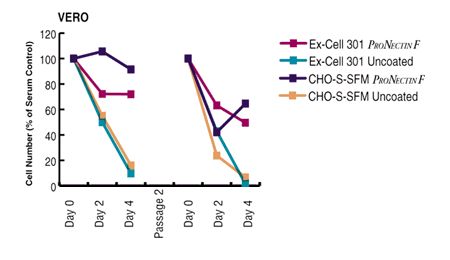
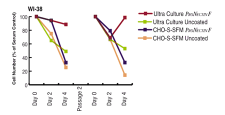
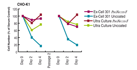
    ProNectin® F consistently outperformed standard tissue culture labware, immediately yielding cell counts equaling 70-100% of the serum control with no prior adaptation4.

    VERO cells in serum-free media
    WI-38 cells in serum-free media

    CHO-K1 cells in serum-free media


    ProNectin F . . . Specific Cell Attachment by Design

    ProNectin® F Diagram ThumbnailProNectin® Fis an engineered recombinant attachment factor which integrates the RGD cell attachment epitope from fibronectin interspersed between structural peptide segments. As shown to the right, the sequence, containing (GAGAGS)9 from silk fibroin, crystallizes in a beta-sheet conformation, where a 10 amino acid sequence from fibronectin containing RGD is presented in a turn, enabling binding to adhesion receptors on the cell surface21,22. Cells are dislodged by standard trypsinization procedures. (To view diagram full size, click on the image.)

    The structural backbone of ProNectin® Fconfers an extraordinary degree of thermal and chemical stability, allowing it to be terminally sterilized and stored for at least one year at room temperature. ProNectin® F provides consistent performance from lot to lot, with no animal-derived contaminants23.


    New! ProNectin Extracellular Matrix Products
    ProNectin F PLUS

    ...a positively charged, water soluble variant of the original ProNectin® F reagent. Combines elements of the functionality of fibronectin, collagen, and polylysine with the stability and ease of use of ProNectin® F.
    ProNectin L

    ...a reagent presenting IKVAV epitopes from the laminin alpha chain.


    References
    (A complete bibliography of references citing use of ProNectin® F is also available.)

    1. Hay, Elizabeth D., editor. 1991. Cell biology of extracellular matrix, 2nd edition.
    2. Ruoslahti, E., Pierschbacher, M.D. 1987. New perspectives in cell adhesion: RGD and integrins. Science 238: 491-497.
    3. Esty, A. 1991. Rapid attachment and spreading of bovine aortic endothelial cells in serum-free and reduced serum media on ProNectin F, an engineered protein polymer. Biomedical Products 16 (5): 76-78.
    4. Putnam, D. and Cappello, J. 1993. Improving the growth of anchorage-dependent cells upon abrupt passage to serum-free media. American Biotechnology Laboratory 11 (13): 14-16.
    5. Varani, J., Inman, D.R., Fligiel, S.E.G., Hillegas, W.J. 1993. Use of recombinant and synthetic peptides as attachment factors for cells on microcarriers. Cytotechnology 13: 89-98.
    6. Gailit, J., Colflesh, D., Rabiner, I., Simone, J., Goligorsky, M.S. 1993. Redistribution and dysfunction of integrins in cultured renal epithelial cells exposed to oxidative stress. American Journal of Physiology 264: F149-F157.
    7. Lwebuga-Mukasa, J.S. 1994. A Mn2+ enhanced, RGD-dependent adhesion technique for isolation of adult rat type II alveolar epithelial cells for immediate functional studies. American Journal of Respiratory Cell and Molecular Biology 10: 347-354.
    8. Wendland, M., Subramani, S. 1993. Cytosol-dependent peroxisomal protein import in a permeabilized cell system. Journal of Cell Biology 120 (3): 675-685.
    9. Waldemar, L., Spear, D. 1993. Improved adherent mammalian cell transfection with ProNectin F recombinant attachment factor. Strategies in molecular biology 5:48-50.
    10. Vedovato, V., Borrow, P., Growing lymphocytic choriomeningitis virus in serum-free media using ProNectin F, an engineered cell attachment polymer. (Manuscript in preparation).
    11. Singhvi, R., Wang, D.I.C., Cappello, J., Production of rIFNg using anchorage-dependent CHO cells cultivated on ProNectin F-coated polystyrene in serum-free medium. (Manuscript in preparation).
    12. Ott, M.J., Olson, J.L., Ballermann, B.J. 1995. Chronic in vitro flow promotes ultrastructural differentiation of endothelial cells. Endothelium 3: 21-30.
    13. Redmond, E.M., Cahill, P.A., Sitzmann, J.V. 1995. Perfused transcapillary smooth muscle and endothelial cell co-culture -- a novel in vitro model. In Vitro Cellular and Developmental Biology -Animal 31: 601-609.
    14. Stanness, K.A., Guatteo, E., Janigro, D. 1996. A dynamic model of the blood-brain barrier In Vitro. Neurotoxicology 17 (2): 481-496.
    15. Davis, T.A., Wiesmann, W., Kidwell, W., Cannon, T., Kerns, L., Serke, C., Delaplaine, T., Pranger, A., Lee, K.P. 1996. Effect of spaceflight on human stem cell hematopoiesis: suppression of erythropoiesis and myelopoiesis. Journal of Leukocyte Biology 60: 69-76.
    16. Yelian, F.D., Yang, Y., Hirata, J.D., Schultz, J.F., Armant, D.R. 1995. Molecular interactions between fibronectin and integrins during mouse blastocyst outgrowth. Molecular Reproduction and Development 41: 435-448.
    17. Lehmann, M., Rabenandrasana, C., Tamura, R., Lissitzky, J.-C., Quaranta, V., Pichon, J., Marvaldi, J. 1994. A monoclonal antibody inhibits adhesion to fibronectin and vitronectin of a colon carcinoma cell line and recognizes the integrins aVß3, aVß5, and aVß6. Cancer Research 54: 2102-2107.
    18. Noiri, E., Gailit, J., Sheth, D., Magazine, H., Gurrath, M., Muller, G., Kessler, H., Goligorsky. M.S. 1994. Cyclic RGD peptides ameliorate ischemic acute renal failure in rats. Kidney International 46: 1050-1058.
    19. Rossi, F., Billetta R., Ruggeri, Z., Zanetti, M. 1995. Engineered idiotypes. Immunochemical analysis of antigenized antibodies expressing a conformationally constrained ARG-GLY-ASP (RGD) motif. Molecular Immunology 32 (5): 341-346.
    20. Ni, J., Chen, S.F., Hollander, D. 1996. Immunological abnormality in C3H/HeJ mice with heritable inflammatory bowel disease. Cellular Immunology 169 (1): 7-15.
    21. Cappello, J., Crissman, J.W. 1990. The design and production of bioactive protein polymers for biomedical applications. ACS Polymer Preprints 31 (1): 193-194.
    22. Anderson, J.P., Cappello, J., Martin, D.C. 1994. Morphology and primary crystal structure of a silk-like protein polymer synthesized by genetically engineered E. Coli bacteria. Biopolymers 34 (8): 1049-1057.
    23. Stedronsky, E.R., Cappello, J., David, S., Donofrio, D.M., McArthur, T., McGrath, K., Panaro, M.A., Putnam, D, Spencer, W., Wallis, O. 1994. Injection Molding of ProNectin F Dispersed in Polystyrene for the Fabrication of Plastic Ware Activated Towards Attachment of Mammalian Cells. Materials Research Society Symposium Proceedings 330: 157-164.
    24. Quinn, P. And Margalit, R. 1996. Beneficial Effects of Co-culture with Cumulus Cells on Blastocyst Formation in a Prospective Trial with Supernumerary Human Embryos. Journal of Assisted Reproduction and Genetics 13 (1): 9-14.


    For Research Use Only.
    U.S. Patent Nos. 5,514,581, 5,211,657, and other patents pending.
    RGD Sequence Licensed from Telios Pharmaceuticals, Inc.


    Site Directory
    webmaster@ppti.com
    Legal Notices

    ©1996-1998 Protein Polymer Technologies, Inc. All rights reserved.
    Redistribution, retransmission, republication or commercial exploitation of the contents of this site
    are expressly prohibited without the written consent of Protein Polymer Technologies, Inc.